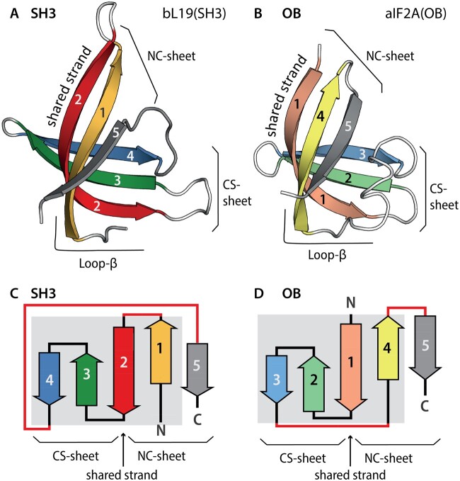
Fig. 1.
The anatomy of SH3 and OB domains. (A) Ribbon and (C) topological representations of the SH3 domain of ribosomal protein bL19 [PDB entry 1VY4, Chain BT]. Strands are colored: β1 yellow; β2 red; β3 green; β4 blue; and β5 gray. (B) Ribbon and (D) topological representations of the OB domain of initiation factor aIF5A [PDB entry 1IZ6, Chain A]. Strands are colored: β1 pink; β2 pale green; β3 light blue; β4 pale yellow; and β5 gray. The shared β-strand, which participates in both β-sheets, is indicated. Differences in strand connectivity are highlighted in red. The common core is indicated by a gray background on the topology diagrams. The consecutive strands sheet (CS-sheet); the N- and the C-terminal β-strands (the NC-sheet).

