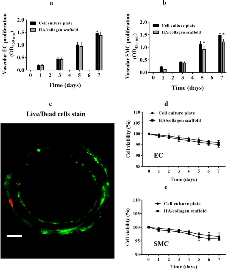
Fig. 3.
The proliferation and viability of vascular ECs and SMCs in the tubular HA/collagen nanofibrous scaffolds. The proliferation of vascular EC in the inner wall (a) and SMC in the outer (b) wall surface of a tubular HA/collagen nanofibrous scaffold and a cell culture plate (positive control) during the period of 7 days (n = 5). *p 0.05. Live/dead stain of ECs and SMCs (c) in a tubular HA/collagen nanofibrous scaffold on day 7 after cell culturing. Scale bars, 500 μm. Quantification of live ECs (d) and SMCs (e) over a period of 7 days (n = 4)

