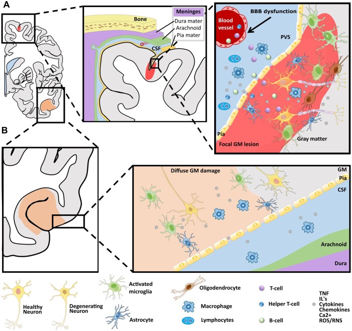
Figure 2.
Simplified schematic depiction of cortical involvement in multiple sclerosis. (A) Focal grey matter lesions (light red) frequently extend along the subpial surface of the cortex. They are topologically related to inflammatory aggregates in adjacent meninges and microglia/macrophages lead to demyelination of the cortex, which is related to a cascade that involves microglial activation and subsequent adaptive immunity via (i) perivascular infiltration of T-cells and B-cells into the GM and/or (ii) infiltration of cytokines, interferon (IFN) and chemokines released by macrophages and other inflammatory cells, such as lymphocytes. (B) Diffuse GM damage (light orange) is mainly related to degeneration of cell bodies and it may be related directly or indirectly to the diffuse microglial activation, which in turn affects trans-synaptic communication leading alterations in network topology. The presence of phagocytic cells may, in first instance, be a reparative process leading to stabilization of the inflammatory response by removal of cellular debris, but it may, however, fail and then lead to incomplete tissue repair. GM = grey matter; WM = white matter; CSF = cerebrospinal fluid; BBB = blood–brain barrier; PVS = perivascular space.

