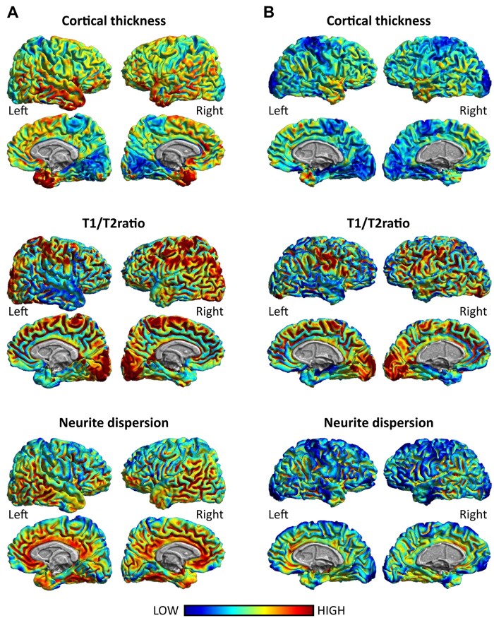
Figure 3.
Example of regional distribution of non-invasive and complementary MRI markers of cortical involvement: cortical thickness, T1/T2 ratio and neurite dispersion. Notice the similarity in the cortical distribution of cortical thickness, T1/T2 ratio and neurite dispersion around the frontal, parietal and temporal cortices across markers. (A) The cortical distribution of these markers in a 36-year-old healthy individual. (B) The cortical distribution of these markers of an age-matched multiple sclerosis patient at disease onset (EDSS: 2; disease duration: 2 years).

