Figure 3.

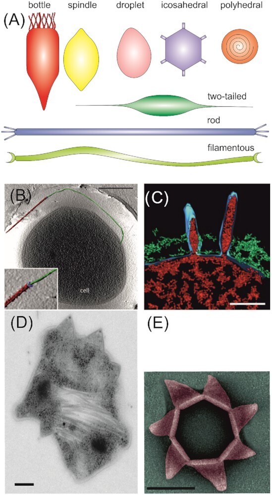
Thermoacidophilic archaeal viruses and their infection mechanisms. (A) Schematic representation of virion morphologies of viruses infecting thermoacidophilic archaea as described in the text. (B) Segmented tomographic volume of an SIRV2 virion (red) attached to a surface filament of Sa. islandicus (green) with help of the three terminal virion fibers (blue). Inset depicts a magnification of the interaction between the tail fibers and the surface structure. Scale bar, 500 nm. (C) Volume segmentations of electron microscopy tomograms showing Sulfolobus spindle-shaped virus 1 maturation and release by budding. Scale bar, 50 nm. (D) Transmission electron micrograph of a thin section of a SIRV2-infected Sa. islandicus cell displaying several pyramidal egress structures. Scale bar, 100 nm. (E)Transmission electron micrographs of an isolated pyramidal egress structure in open conformation isolated after SIRV2 infection of Sa. islandicus. Scale bar, 100 nm. Adapted from Bize et al. (2009); Quax et al. (2011); Quemin et al. (2013, 2016).
