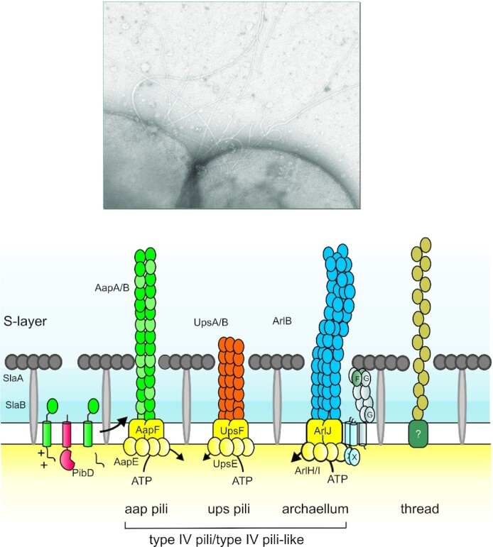
Figure 7.

Archaeal cell surface structures involved in planktonic and biofilm growth. Upper image: electronic microscopy image from a S. acidocaldarius cell where archaellum and pilus can be seen. The upper image is reproduced from Albers and Meyer 2011. Lower image: schematic model with all proposed cell surface appendages in the Sulfolobales: the Aap pili (archaeal adhesive pili), the Ups pili (UV-induced pili), the archaellum and the threads. Also depicted is the S-layer and its proteins, SlaA and SlaB.
