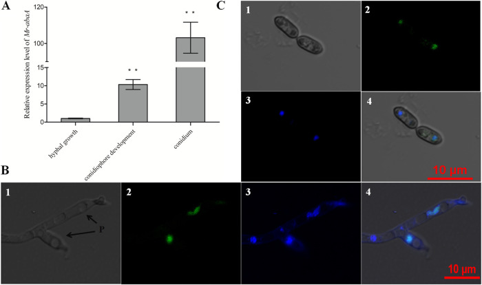
FIG 2.
Transcriptional profiles and subcellular localization of Mr-AbaA. (A) Relative transcript levels of Mr-abaA in the WT cultures in three different developmental stages, compared with the standard level during hyphal growth. **, P < 0.01, Tukey’s HSD tests. (B and C) Subcellular localization of AbaA::GFP fusion protein expressed in the conidiophore development (B) and conidium (C) stages. Nuclei were stained with DAPI. Brightfield, expressed (green), DAPI-stained (blue), and merged views of the same field are numbered 1, 2, 3, and 4, respectively. P, phialides. Error bars in panel A indicate standard deviations of the means from three independent replicates.

