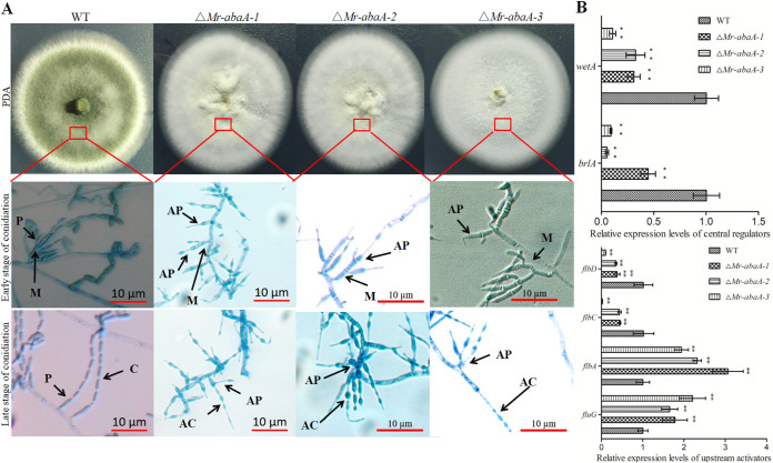
FIG 3.
Phenotypic analysis of the WT and ΔMr-abaA strains. (A) Three independent ΔMr-abaA strains showed a change in colony color. To observe the conidiophores on the aerial hyphae, WT and ΔMr-abaA cells were grown on PDA plates and sampled at 2.5 dpi (early stage of conidiation) and 10 dpi (late stage of conidiation). WT conidiophores had metulae (M), phialides (P), and conidia (C), whereas ΔMr-abaA conidiophores had metulae (M), abnormal phialides (AP), and abacus aberrant conidia (AC). (B) qRT-PCR analysis of the expression levels of conidiation-related genes among WT and null mutant clones. **, P < 0.01, Tukey’s HSD tests. Error bars in panel B indicate standard deviations of the means from three independent replicates.

