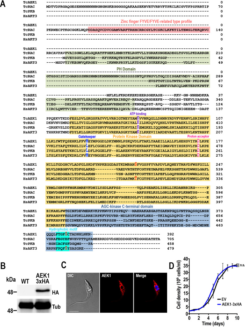
FIG 1.
TcAEK1 structure and localization. (A) Amino acid sequence alignment of AGC kinase subfamily members from T. cruzi, TcAEK1, TcRAC, and TcPKB (TriTrypDB IDs: TcYC6_0120630, TcYC6_0106530, TcYC6_0095770, respectively), and human HsAKT3 (GenBank accession number: NP_001357003). Important conserved domains and residues are highlighted. In red are putative conserved phosphorylated amino acid residues. PH, Pleckstrin homology domain. (B) TcAEK1-3×HA (AEK1-3×HA) overexpression was confirmed by Western blotting using anti-HA antibodies. Cells transfected with pTREX-n empty vector (EV) were used as the control cell line. Tubulin (Tub) was used as the loading control. (C) IFA showed subcellular localization of TcAEK1-3×HA detected with anti-HA antibodies (AEK1 [red]) in T. cruzi epimastigotes. Merge of red signal and DAPI staining (blue) and DIC (differential interference contrast) images is also shown. Scale bar is 5 μm. (D) Growth of control (EV) and TcAEK1 (AEK1-3×HA) epimastigotes in LIT medium. No significant (n.s.) differences in growth rates were found using one-way ANOVA with multiple comparisons (n = 3).

