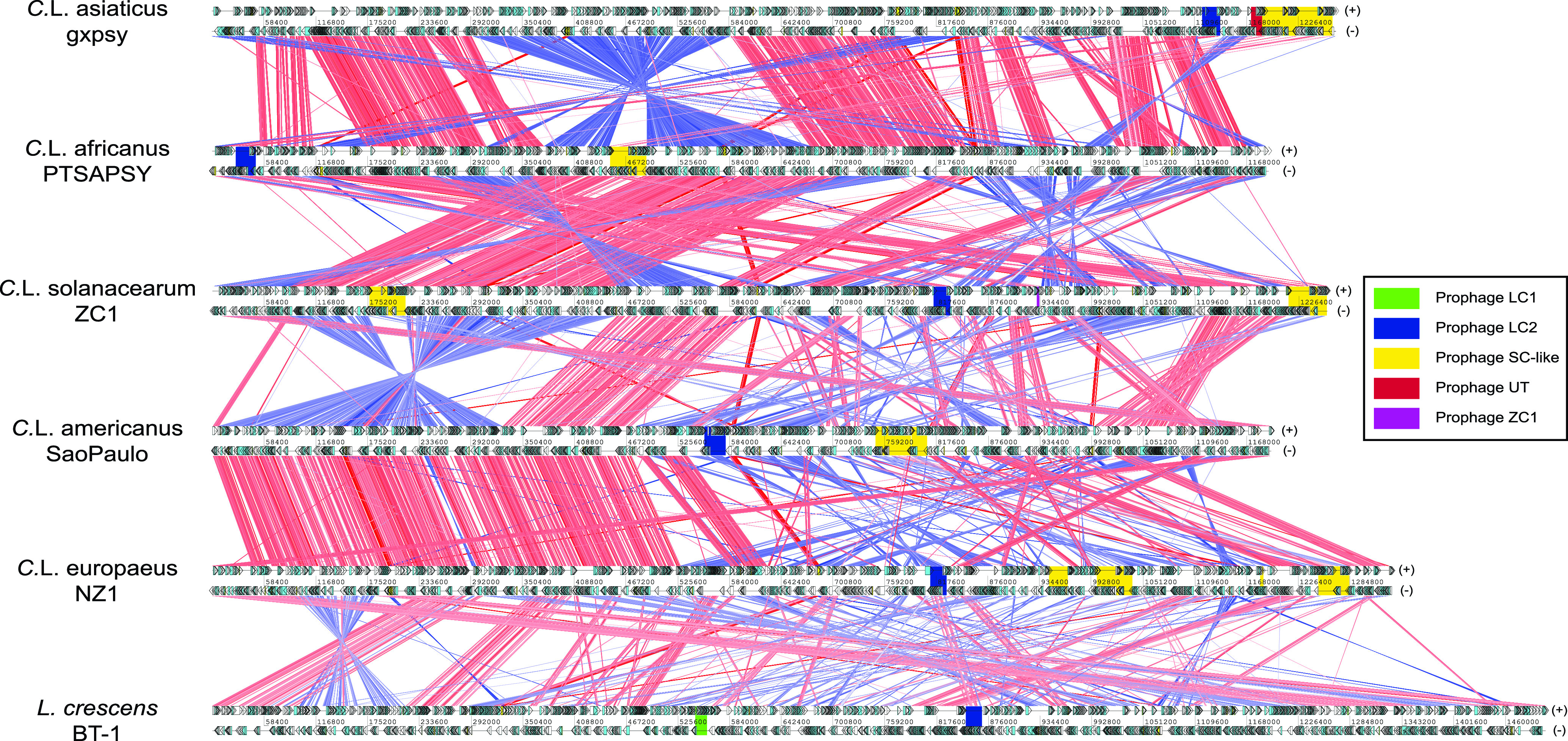
FIG 2.

Whole-genome comparisons of Liberibacter species. The genomes are labeled by their scientific names followed by strain information. Cyan and yellow boxes are the protein coding frames and RNA regions on either forward (+, top) or reverse (−, bottom) strands, respectively. Homologous genes between genomes are linked by lines, where the red and blue lines represent the forward and reverse (complementary) matches, respectively. The intensity of the color bands is proportional to the percent identity of the match, where higher intensity indicates higher sequence identity. The phage loci are shown in colored boxes (legend on the right).
