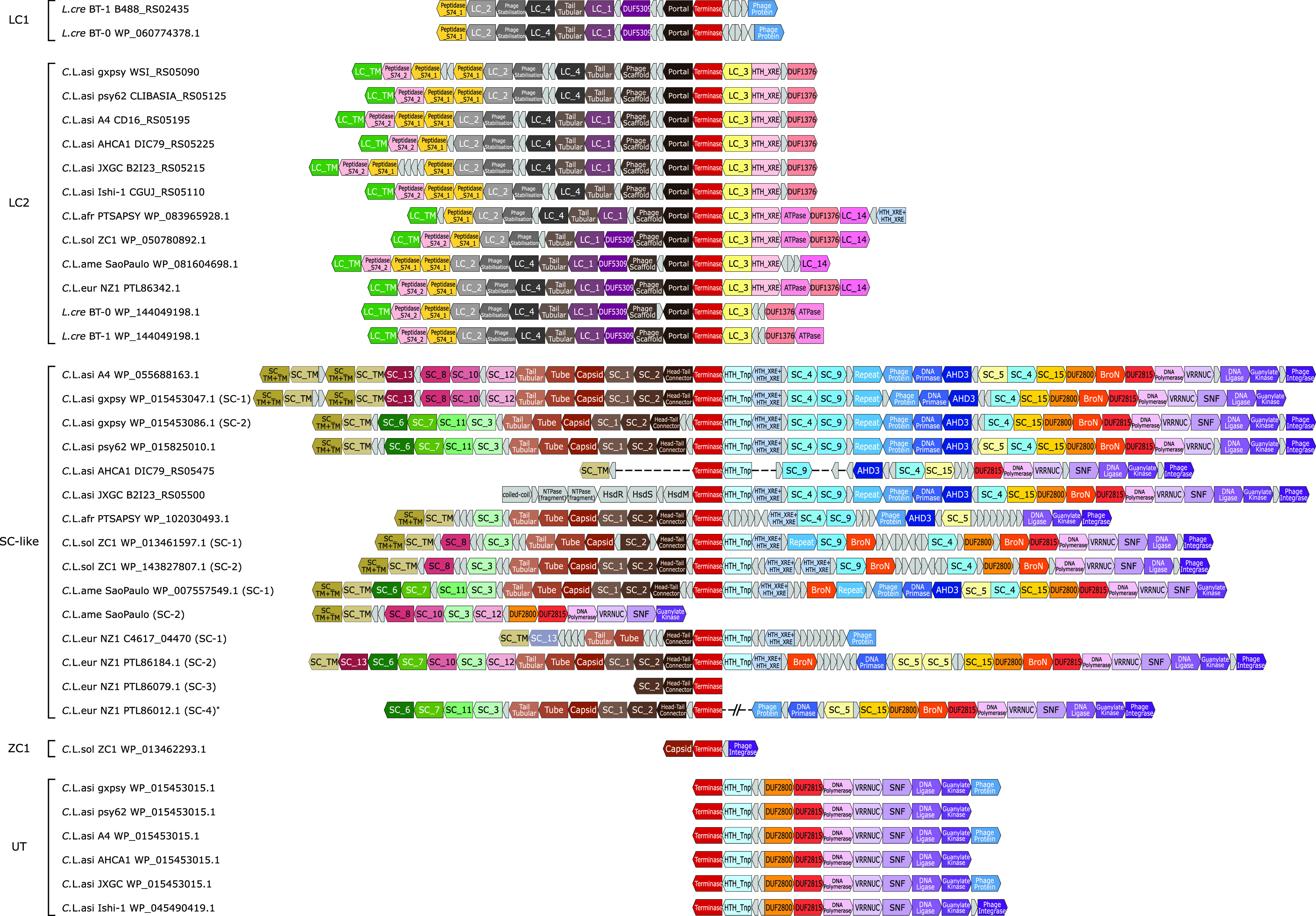
FIG 3.

Genomic structures of the prophages identified in the Liberibacter species. The coding frames in prophage loci are presented in blocks. The blocks, labeled with gene annotations and highlighted in different colors, are the coding products shared by at least four prophage loci, while the small gray blocks represent nonconserved coding products or pseudogenes. The genome structures were aligned based on the shared terminases of these prophage loci. On the left side, the prophage loci are indicated by their species names, strains, and terminase accession numbers or locus tags. The prophage loci were classified into four types (LC1, LC2, SC-like, UT) and a distinct prophage fragment found in a “Ca. Liberibacter solanacearum” ZC1 strain. One “Ca. Liberibacter europaeus” SC-like prophage structure was derived from two genome contigs, PSQJ01000015.1 and PSQJ01000003.1, which is indicated by an asterisk (*).
