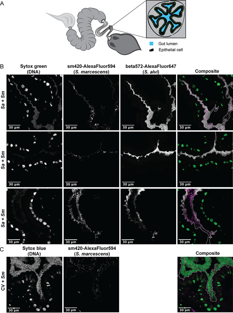
FIG 2.
Gut commensals compete with Serratia marcescens for space. (A) Diagram of the honey bee gut, showing a cross-section of the ileum. (B) Representative cross-sections of ilea from bees colonized by S. alvi (white) and S. marcescens (magenta). MF bees were inoculated with S. alvi wkB2 and exposed to S. marcescens after 5 days. S. alvi and S. marcescens were visualized using fluorescent probes that hybridize to 16S rRNA, while Sytox green stain indicates the presence of host nuclei and bacterial cells (green). (C) Cross-section of the ileum of a bee inoculated with gut homogenate (CV) and then exposed to S. marcescens (magenta). Sytox blue stain was used to label host nuclei and bacterial cells (green). Dashed white lines on the composite images outline the lumen of the gut.

