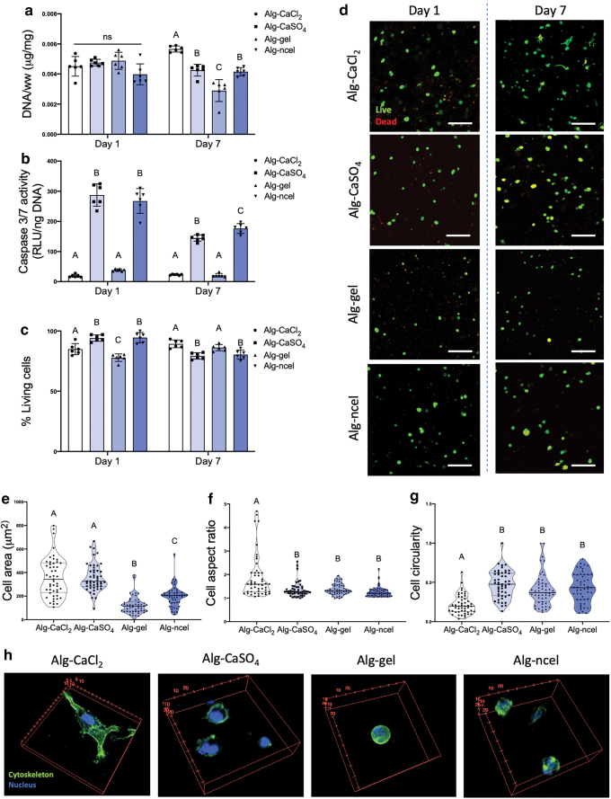
FIG. 5.
MSC viability, metabolic activity, and cell–substrate interaction depend on the type of alginate-based bioink. (a) DNA content in 3D printed constructs at days 1 and 7 after biofabrication. (b) Caspase 3/7 activity in the 3D printed constructs at days 1 and 7 after biofabrication. (c) Percentage of living cells in the 3D printed gels at days 1 and 7 after printing (n = 6 for panels a–c). (d) Live/dead confocal images of cells in 3D printed gels at days 1 and 7 after printing (live cells are green, dead cells are red). Quantification of (e) cell area, (f) cell aspect ratio, and (g) cell circularity as shape descriptors of cells encapsulated in 3D printed cells at day 7 after printing (n = 49 for panels e–g; ImageJ was used to analyze three images per group). (h) 3D confocal reconstructions of cells encapsulated in 3D printed constructs and stained for their actin cytoskeleton (green) and nucleus (blue). Scale bar = 200 mm. Color images are available online.

