Fig. 2.
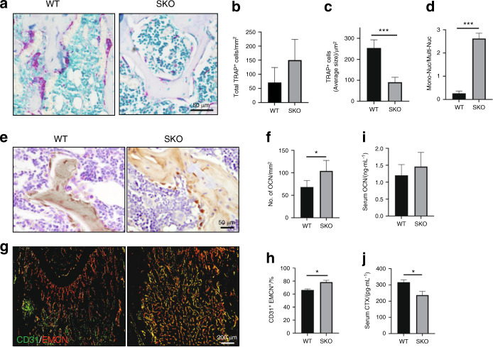
The numbers of mononuclear TRAP+ cells, type H vessels, and osteoblasts were increased in the SKO mice. a Representative images of TRAP staining of the distal femurs of 3-month-old WT and SKO mice. b–d Quantitative analysis of the TRAP staining in a. The number and size of TRAP+ cells were normalized to the bone area. Mon-Nuc: mononuclear TRAP+ cells, Multi-Nuc: multinuclear TRAP+ cells. n = 5, ***P < 0.001. Error bar represents the standard error of the mean. e Representative images of immunohistological staining of osteocalcin in the distal femurs of the 3-month-old WT and SKO mice. f Quantitative analysis of the osteocalcin staining in e. n = 5, *P < 0.05. Error bar represents the standard error of the mean. g Cofluorescence staining of CD31 (green) and endomucin (EMCN) (red) in mouse femurs. h Quantitative analysis of the ratio of CD31 and EMCN double-positive fluorescence intensity in g. n = 5, *P < 0.05. Error bar represents the standard error of the mean. ELISAs of OCN (i) and CTX (j) in the serum of the WT and SKO mice. The mice were sacrificed at 3 months of age. n = 5, *P < 0.05

