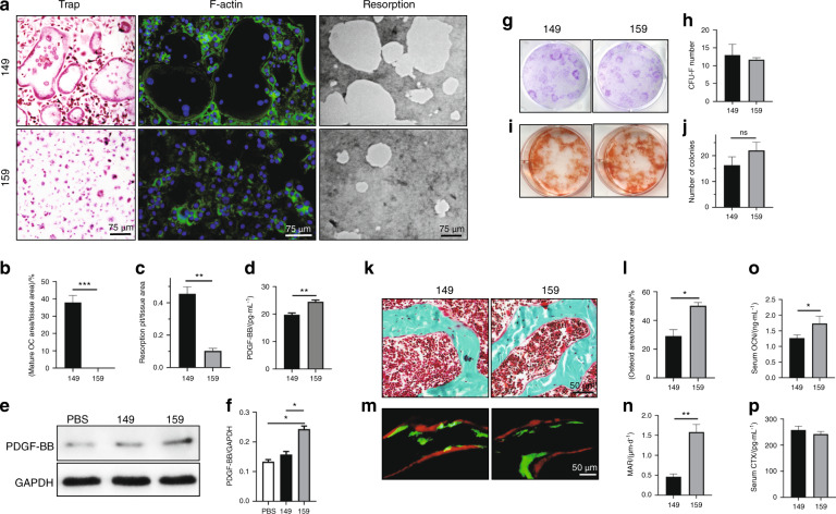
Fig. 4.
Injection of a Siglec-15 neutralizing antibody halted the maturation of osteoclasts and increased bone formation. a Representative images of TRAP staining (first column), phalloidin staining (second column), and resorption pits (third column) of the osteoclast cultures that were induced from primary mouse bone marrow monocytes. All experiments were conducted on day 7 of osteoclastogenic induction. b, c Quantitative analysis of the TRAP staining and resorption area in a. **P < 0.01, ***P < 0.001. d ELISAs of the concentration of PDGF-BB in the conditioned media that was collected on day 7 of osteoclastogenesis. **P < 0.01. e Western blot of PDGF-BB in the cell lysate of osteoclasts on day 7 of osteoclastogenic induction. f Quantitative analysis of blot size in e. *P < 0.05. g, h Crystal violet staining and quantitative analysis of bone marrow stromal cells cultured for 10 days. i, j Alizarin red staining of bone marrow stromal cells cultured in osteogenic medium for 21 days. Bone marrow stromal cells were collected from 4-month-old mice that received NP-149 or NP-159 injection for 1 month. ns: not significant. k Trichrome staining of femurs collected from the mice treated with NP-149 or NP-159 for 2 months. The osteoid layer is labeled red. l Quantitative analysis of the osteoid-covered surface. n = 5, *P < 0.05. m Double labeling of the mineral layers in mouse femur trabecular bone. The mice were treated with NP-149 or NP-159 for 1 months. n Quantitative analysis of the mineral apposition rate in m. n = 5, **P < 0.01. ELISAs of OCN (o) and CTX (p) in the serum from the mice treated with NP-149 or NP-159 for 1 month. n = 5, *P < 0.05

