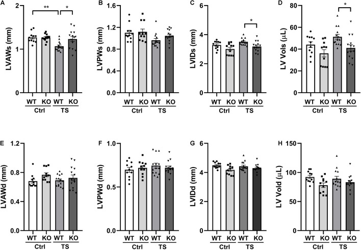
FIGURE 3.
The left ventricular structure of WT and WWP1 KO mice after tail suspension. (A–H) Quantitative analysis of the diastolic and systolic left ventricular posterior wall thickness (LVPWd and LVPWs), LV anterior wall thickness (LVAWd and LVAWs), LV internal diameter (LVIDd and LVIDs), and LV volume (LV Vold and LV Vols) from WT and WWP1 KO mice by echocardiography following tail suspension. Data represent the means ± SEM. *P < 0.05, **P < 0.01. Ctrl, control; TS, tail suspension; WT, wild-type mice; KO, knockout.

