FIGURE 5.
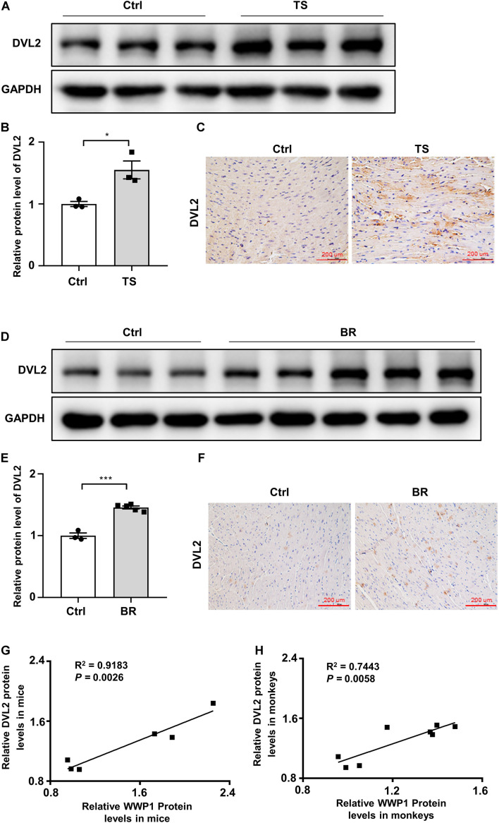
Disheveled segment polarity proteins 2 expression changes in the hearts of mice and rhesus monkeys after simulated microgravity. (A) Representative western blotting analysis of DVL2 expression in cardiac extracts of adult mice following tail suspension (TS) for 6 weeks (n = 3 for each group). (B) Quantification of DVL2 protein levels of (A). (C) Immunohistochemistry for DVL2 (brown) in paraffin section from mouse hearts at 6 weeks after tail suspension. Scale bars, 200 μm. (D) Representative western blotting analysis of DVL2 expression in cardiac extracts of adult rhesus monkeys following bed rest (BR) for 6 weeks (Ctrl, n = 3; BR, n = 5). (E) Quantification of DVL2 protein levels of (D). (F) Immunohistochemistry for DVL2 (brown) in a paraffin section from rhesus monkey hearts at 6 weeks after bed rest. (G) Pearson correlation coefficients between DVL2 and WWP1 protein levels in the heart of mice following tail suspension. (H) Pearson correlation coefficients between DVL2 and WWP1 protein levels in the heart of rhesus monkeys following bed rest. Scale bars, 200 μm. Data represent the means ± SEM. *P < 0.05, ***P < 0.001. DVL2, disheveled segment polarity protein 2; TS, tail suspension; BR, bed rest.

