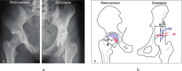
Fig. 1.
a) Radiological and b) schematic views of young patients with hip pain due to acetabular retroversion (left) and hip dysplasia (right). b) Three radiological signs of acetabular retroversion are shown (positive ischial spine sign, posterior wall sign and crossover sign with retroversion index of >50%) . AI, acetabular index; LCE, lateral-centre edge.

