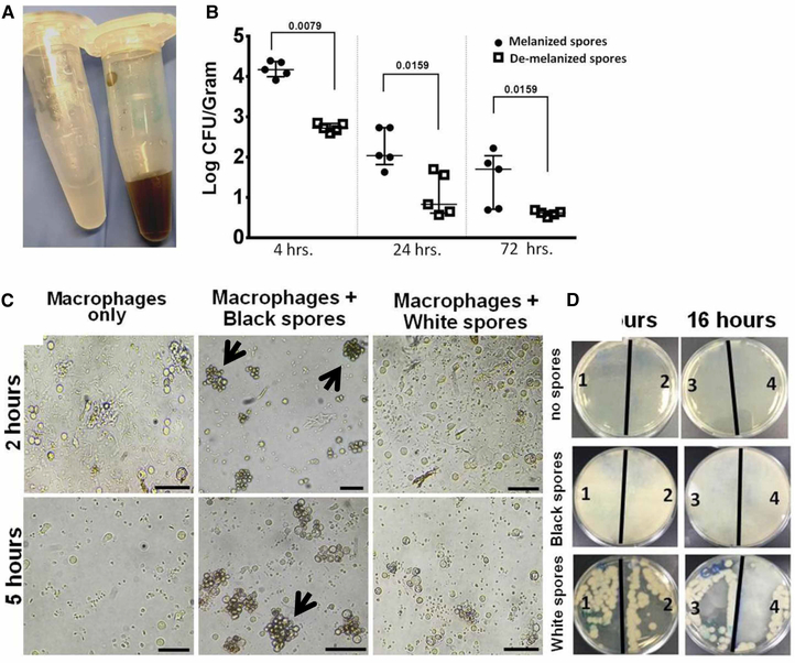
Figure 10. De-melanization of Rhizopus (white) spores enhanced the phagocytic ability of macrophages.
(A) White Rhizopus spores developed after treatment with UOSC-2 for 3 days compared with wild-type black spores. (B) Fungal burden from the lungs of infected immunocompetent mice (n = 5 per experimental group) with 106 fungal spores. The mice were sacrificed at the indicated time points, lungs were collected and the lungs fungal burden were assessed by CFU plating on PDA containing 0.1% triton. The data were analyzed using Student’s t-test and statistical significance was calculated. The data display the mean ± SEM of five different mice. (C) Representative photomicrographs of macrophages obtained from peripheral blood monocytes incubated with Rhizopus spores at a ratio of 1 macrophage to 2 spores at different time points. Developed white spores failed to cluster and were eliminated rapidly as compared with non-treated wild-type spores (indicated by black arrows). Scale bar = 50 μm. (D) Viability of spores after phagocytosis events. At the end of each time point, excess spores were washed with pre-warmed PBS and the remaining macrophages were lysed. The lysates were further diluted and cultured overnight on PDA. White spores viable numbers capable of growing on media were reduced after phagocytosis when compared with wild-type spores. The numbers on the plates (1–4) indicated the number of replicas per each treatment.

