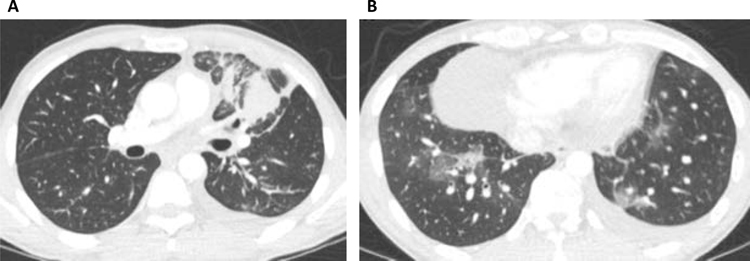
Figure 3. Lymphangitic carcinomatosis associated with ALK+ lung cancer.
Representative axial CT images are taken from a 43-year-old male never smoker who presented with (A) dominant left upper lobe mass with surrounding lymphangitic carcinomatosis and (B) additional areas of lymphangitic carcinomatosis in the lower lobes.

