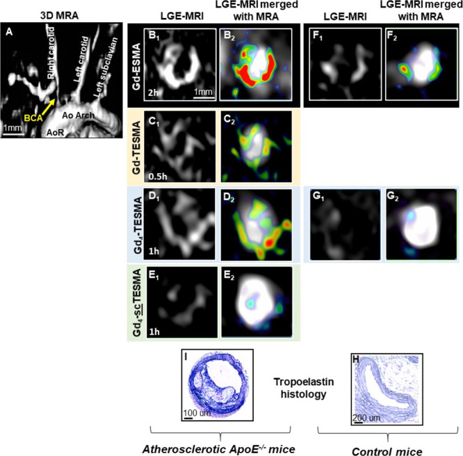
Figure 7.

Molecular imaging of tropoelastin in atherosclerotic ApoE–/– and control mice using the tetrameric Gd4-TESMA contrast agent. (A) The 3D reconstructed angiogram (MRA) shows the vasculature. Late gadolinium enhancement (LGE) images (B1–D1) and merged LGE and angiography images (B2–D2) compare the signal enhancement of atherosclerotic plaques located in the brachiocephalic artery of the same animal imaged serially after the administration of the elastin-binding Gd-ESMA (2 h), the tropoelastin-binding Gd-TESMA (0.5 h), and the tropoelastin-binding tetrameric Gd4-TESMA (1 h) agents. (E1, E2) LGE and merged images show a low uptake of the tetrameric tropoelastin-binding agent attached to a scrambled peptide (Gd4-scTESMA). (F1, F2) LGE and merged LGE and angiography images show signal enhancement of the brachiocephalic artery of control mice after the injection of the elastin agent Gd-ESMA but (G1, G2) no enhancement after the injection of the tropoelastin agent Gd4-TESMA. Immunohistochemistry using a tropoelastin antibody shows the absence of tropoelastin in control tissues (H) and dense tropoelastin molecules (dark/purple signal) in the disease artery (I).
