Fig. 2.

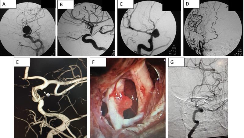
Preoperative and postoperative angiograms of various ophthalmic segment aneurysms. ( A, B ) Dorsal wall, ( C, D ) Ventral wall, ( E, G ) Blister aneurysm, and (F) shows intraoperative picture of a blister aneurysm.

Preoperative and postoperative angiograms of various ophthalmic segment aneurysms. ( A, B ) Dorsal wall, ( C, D ) Ventral wall, ( E, G ) Blister aneurysm, and (F) shows intraoperative picture of a blister aneurysm.