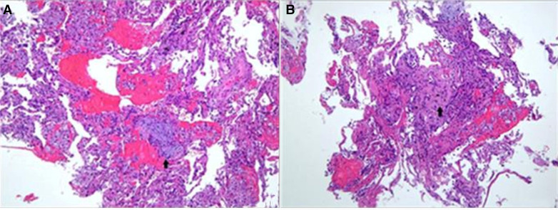
Figure 2.
(A) Fragments are composed of benign pneumocytes lining the alveolar spaces, with scattered foamy macrophages, haemorrhage and occasional fibroblastic foci within the space (arrow). H&E, ×100. (B) The alveolar septa are focally fibrotic (arrow) and there are mild to moderate inflammatory cell infiltration within the stroma. H&E, ×100.

