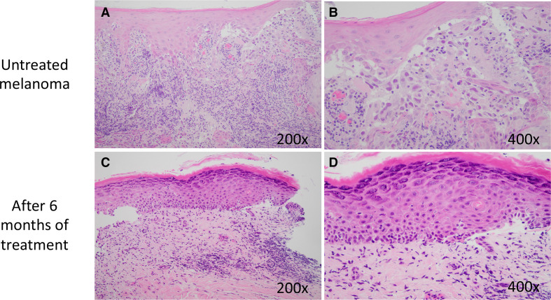
Figure 2.
Photomicrographs of the untreated lesion (A) show recurrent malignant melanoma arising from surface epithelium directly invading into the underlying lamina propria. Higher power (B) shows tumor cells with nuclear pleomorphism and melanin granules within the cytoplasm. After 6 months of treatment, histology shows surface epithelium with hyperkeratosis and underlying chronic inflammatory infiltrate (C). There is no evidence of residual melanoma at higher magnification (D).

