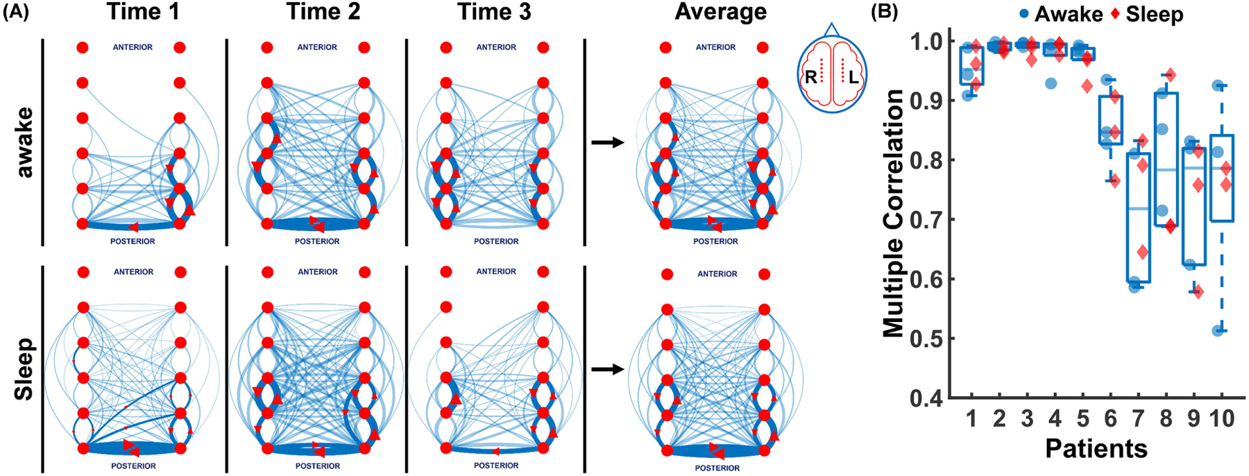
Figure 2. Interictal spike networks are highly reproducible and independent of sleep wake cycles.

(A) The top 10% (solid blue lines with red arrow marks) and 90% remaining (faint blue lines) propagations are shown for patient 8 at three different awake and sleep 10 min intervals (Time1, Time2, Time3). Propagations here include all EEG activity between the broad band of 1–35 Hz. The average of the awake and sleep propagations are shown on the right panel which shows the high level of similarity for both wake and sleep spike propagations. (B) Multiple correlation coefficients across 3 sleep (red diamonds) and 3 awake (blue circles) 10 min EEG segments for each of 10 patients (0.93±0.13) is shown demonstrating the high reproducibility of the measured spike networks in both wake and sleep states.
