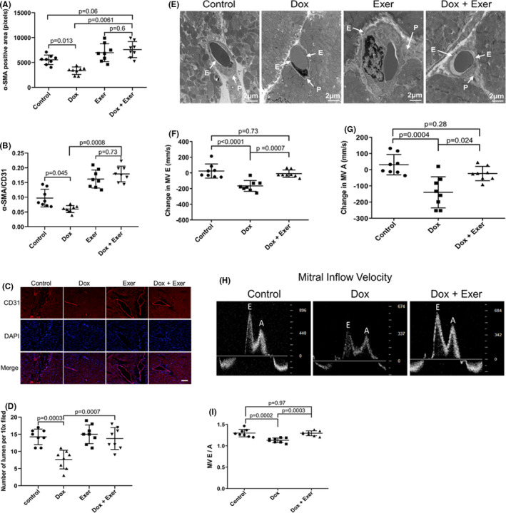
FIGURE 3.

Effect of doxorubicin with and without exercise on cardiac vessel morphology and cardiac diastolic blood flow in nude mice following Dox therapy. (A) Quantitative analysis of the total area of pericytes (as defined by α‐SMA) in the heart tissues. (B) Ratio of α‐SMA/CD31 cells (mean ± SD for five random fields). (C) Representative images of CD31+ staining. Scale bar: 50 μm. (D) Quantitative analysis of number of open lumens in vessels >100 μm from each treatment group (mean ± SD; Control versus Dox, p = 0.0003; Dox versus Dox + Exer, p = 0.0007). Statistical analysis as described in Figure 2. (E) Representative transmission electron microscopy images of cardiac vessels. Vascular endothelial cells and pericytes are indicated by the arrow. E indicates endothelial. P indicates pericytes. (F, G) Change in mitral valve E and A velocity 2 weeks after therapy. (H) Representative echocardiography images of the E and A peak. (I) E/A ratio was measured for each mouse. Dox indicates doxorubicin and Exer indicates exercise
