
FIGURE 5.
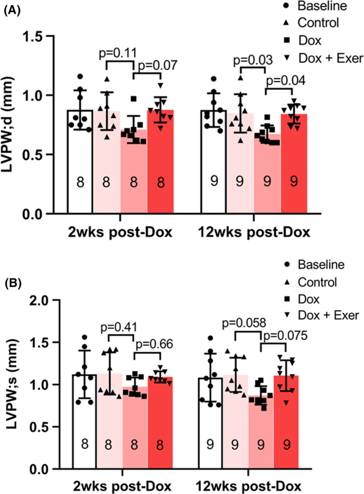
Effect of doxorubicin with or without exercise on diastolic and systolic function in nude mice 12 weeks after therapy. Diastolic (A) and systolic (B) functions were assessed after therapy and 12 weeks later using echocardiography by measuring left ventricular posterior wall thickness in diastole (LVPW; d) and systole (LVPW; s). At 2 weeks post‐Dox (n = 8 mice per group), control versus Dox, p = 0.11; Dox versus Dox + Exer, p = 0.07; at 12 weeks post‐Dox (n = 9 mice per group), control versus Dox, p = 0.03; Dox versus Dox + Exer, p = 0.04. Statistical analysis as described in Figure 2