
FIGURE 8.
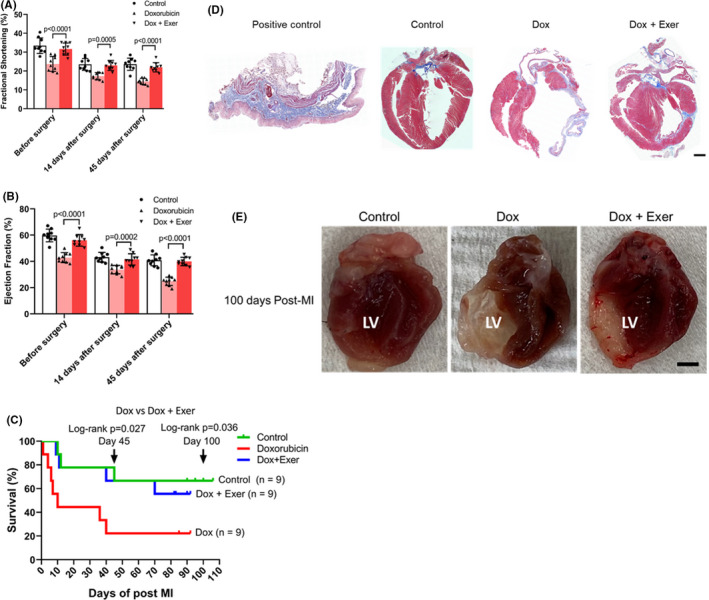
Recovery following left anterior descending artery ligation (myocardial infarction). Myocardial infarction (MI) surgery was performed 90 days after treatment with doxorubicin (Dox), Dox + exercise (Exer), or no therapy (control). (A, B) Fractional shortening (FS) and ejection fraction (EF) were assessed before surgery and then 14 and 45 days after surgery. ****p < 0.0001. Statistical analysis as described in Figure 2. (C) Survival rates were quantified following MI for each group. Kaplan–Meier survival curves of mice at day 45 and day 100 (arrows) after MI. n indicates number of mice. *p < 0.05. (D) Collagen deposition in the left ventricle was assessed using Masson trichrome staining 100 days after MI. Mice treated with Dox alone had a thinned left ventricle with collagen deposition in the left ventricular wall. This was not observed in the mice treated with Dox + Exer. Scale bar: 0.5 mm. (n = 9 in each group). (E) Left ventricular wall heart tissue following myocardial infarction (MI) for 100 days in nude mice. LV indicates left ventricle. Dox indicates doxorubicin. Exer indicates exercise