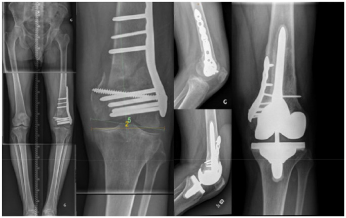
Fig. 11.

This composite figure illustrates a left KA TKA performed on a post-traumatic knee. The patient sustained a complex articular distal femoral fracture that was treated with ORIF, and developed a medial femoral condyle malunion (substantial proximal migration). The right knee anatomy (LDFA 3.5° valgus and MPTA 5° varus) was used as a template for planning the left KA TKA. To balance the left KA TKA, an osteotomy of the medial femoral condyle had to be performed in order to correct the malunion. Postoperative radiograph shows a left TKA with LDFA of 3° valgus and MPTA of 5° varus. A 100 mm loose fit 12 mm cemented femoral stem was used to supplement femoral fixation and protect the medial condyle fixation.
Note. KA, Kinematic Alignment; TKA, total knee arthroplasty; ORIF, open reduction and internal fixation; LDFA, lateral distal femoral angle; MPTA, medial proximal tibial angle.
