
Fig. 2.
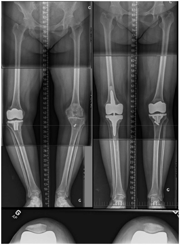
This composite figure illustrates a 65-year-old female patient with constitutional ‘Type 1’ left knee. The patient undertook a right MA TKA that failed after four years. On the left knee, the native lateral distal femoral angle (LDFA) was 2 degrees valgus and the medial proximal tibial angle (MPTA) 9 degrees varus. In the same setup, a left computational assisted rKA TKA and a right TKA revision were performed. rKA boundaries were applied when implanting the left knee: the tibial varus was reduced to 5 degrees with a resulting HKA of 3 degrees varus. Deep MCL had to be released to obtain medio-lateral compartment balance.
Note. MA, mechanical alignment; TKA, total knee arthroplasty; rKA, restricted kinematic alignment; HKA, hip-knee-ankle; MCL, medial collateral ligament.