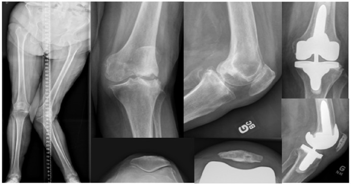
Fig. 8.
This composite figure illustrates a 79-year-old woman with a severe ‘Type 5’ valgus left knee. The patient developed a severe deformity secondary to avascular necrosis of her lateral femoral condyle. There was substantial bipolar (femoral and tibial) lateral compartment bone loss and a stretched MCL. The right knee anatomy was used to plan the left knee TKA. Using computer navigation, the left femoral implant was aligned with 5° of valgus and the tibial component with 2° of varus. A 10 mm lateral femoral augment was used to fill the bone defect and a short-cemented stem for supplementary fixation. A postero-stabilized semi-constrained insert was used to compensate for the stretched MCL.
Note. MCL, medial collateral ligament; TKA, total knee arthroplasty.

