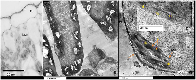
Figure 11.
Anatomical structure of Tomato leaf cv “Super marmende”growing under stress chilling conditions 5 days/4ºC for 6h/day treated with GABA at 2mM. (A) Leaf cross-section showed the upper epidermis (Ep) and the two mesophyll layers (Mes); note that chloroplasts were accumulated together in clusters. (B) TEM micrograph showed mesophyll ultra-structures: There are expansions in the size of the plastids due to the large starch grains in them, but with a tendency to elongate shape. (C,D) high magnification of one chloroplast showed normal chloroplast membrane (ChM), well-developed grana (G), and stromal thylakoids (Th), the cell wall (CW) was attached well with the outer cellular membrane.

