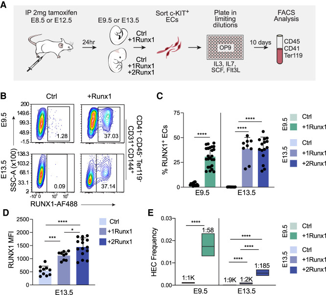
Figure 1.
RUNX1 can efficiently specify both embryonic and fetal ECs as hemogenic, although fetal ECs require a higher dose of RUNX1. (A) Schematic illustrating limiting dilution assays for quantifying HEC frequency. (B) Representative contour plots of RUNX1 expression in ECs from Ctrl and +Runx1 embryos and fetuses 24 h after tamoxifen delivery. (C) Quantification of percentage (mean ± SD) of RUNX1+ ECs. (D) Quantification of the median fluorescence intensity (MFI; ±SD) of RUNX1 expression in ECs from E13.5 Ctrl, +1Runx1, and +2Runx1 fetuses. For B–D, ECs were purified as CD41−CD45−Ter119−CD144+CD31+ cells (Supplemental Fig. S1B). Each point represents pooled embryos (E9.5) or a single fetus (E13.5). Data are from three E9.5 and six E13.5 litters/independent experiment. To determine significance, unpaired two-tailed Student's t-test was used for E9.5 and one-way ANOVA and Tukey's multiple comparison test was used for E13.5. (E) Frequency of HECs (± 95% CI) in c-KIT+ ECs. HEC frequencies are indicated above the bars. Data represent four biological replicates using pooled cells from litters of E9.5 embryos collected in four independent experiments, and three biological replicates using pooled cells from E13.5 fetuses collected in three independent experiments. Significance was determined using ELDA. In all figures: (****) P ≤ 0.0001, (***) P ≤ 0.001, (**) P ≤ 0.01, (*) P ≤ 0.05, (ns) P > 0.05.

