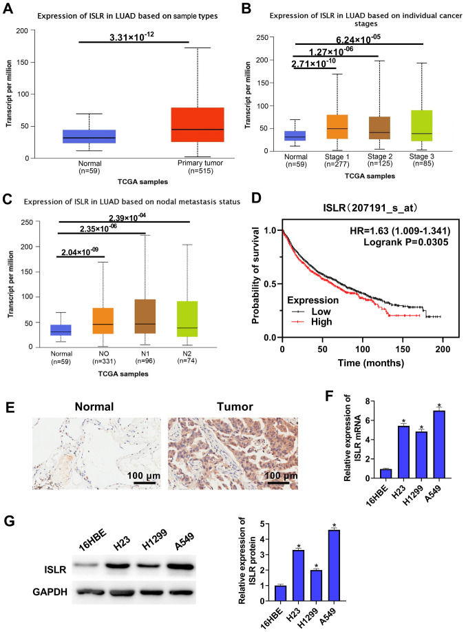
Figure 1.
ISLR expression is elevated both in NSCLC tissues and cell lines. (A) TCGA-LUAD cohort datasets showed that ISLR expression was higher in tumour tissues than in normal tissues. Analysis of TCGA data showed that ISLR was highly expressed in cancers at (B) different stages and (C) in nodal metastasis cancer. (D) Kaplan-Meier survival plots demonstrated that higher ISLR abundance correlated with poorer OS, as determined using TCGA database. (E) ISLR expression in non-tumorous lung tissues and primary lung cancer tissues was measured via immunohistochemistry. Scale bar, 100 µm. ISLR expression in NSCLC cell lines (A549, H1299 and H23) and normal immortalized lung epithelial cell lines (16HBE) was measured via (F) reverse transcription-quantitative PCR and (G) western blotting. *P<0.05 vs. 16HBE cells. NSCLC, non-small cell lung cancer; TCGA, The Cancer Genome Atlas; ISLR, immunoglobulin superfamily containing leucine-rich repeat; LUAD, lung adenocarcinoma.

