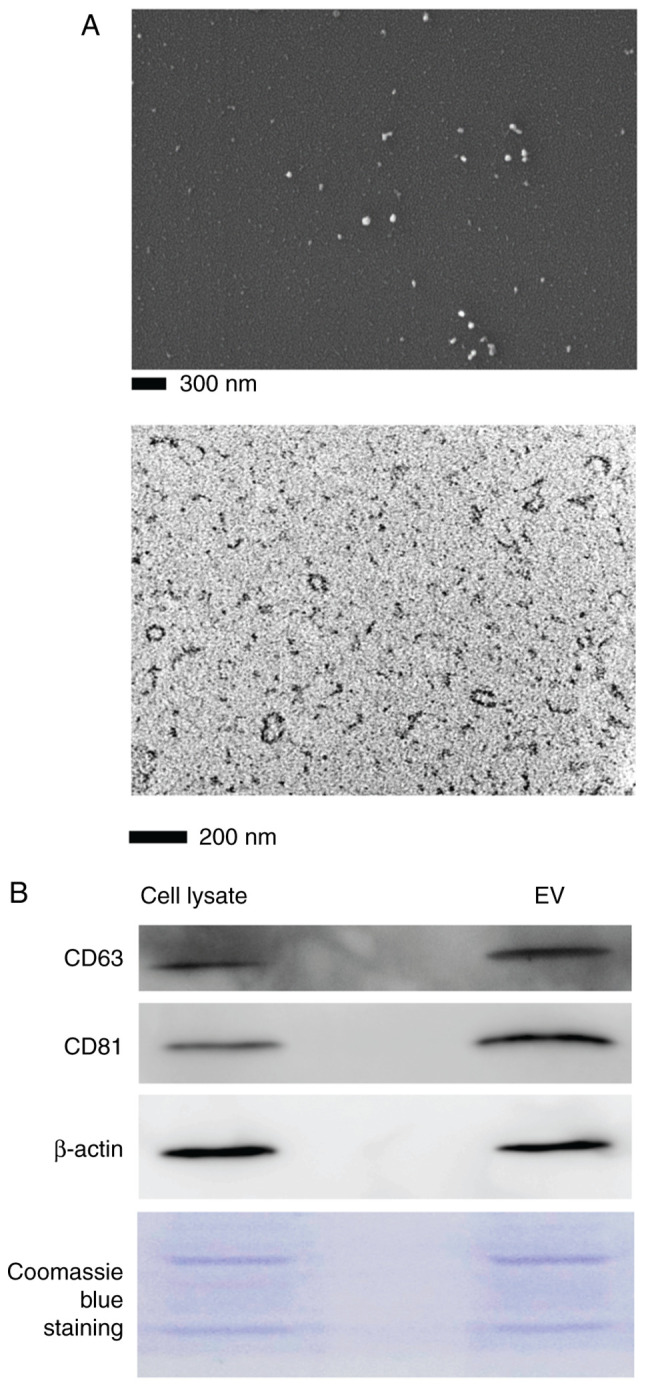
Figure 2.

Characterization of T-MSC EVs. (A) Scanning (upper; magnification, ×70,000) and transmission electron microscopy (lower; magnification, ×200,000) images of EVs released from T-MSCs. (B) Immunoblot analysis of EV specific markers, CD63 (26 kDa) and CD81 (22-26 kDa), from cell lysates and EVs of T-MSCs. β-actin (42 kDa) and Coomassie blue staining gel were used as a loading control (5 µg/lane). T-MSC, tonsil-derived mesenchymal stem cell; EV, extracellular vesicle.
