
Fig. 7.
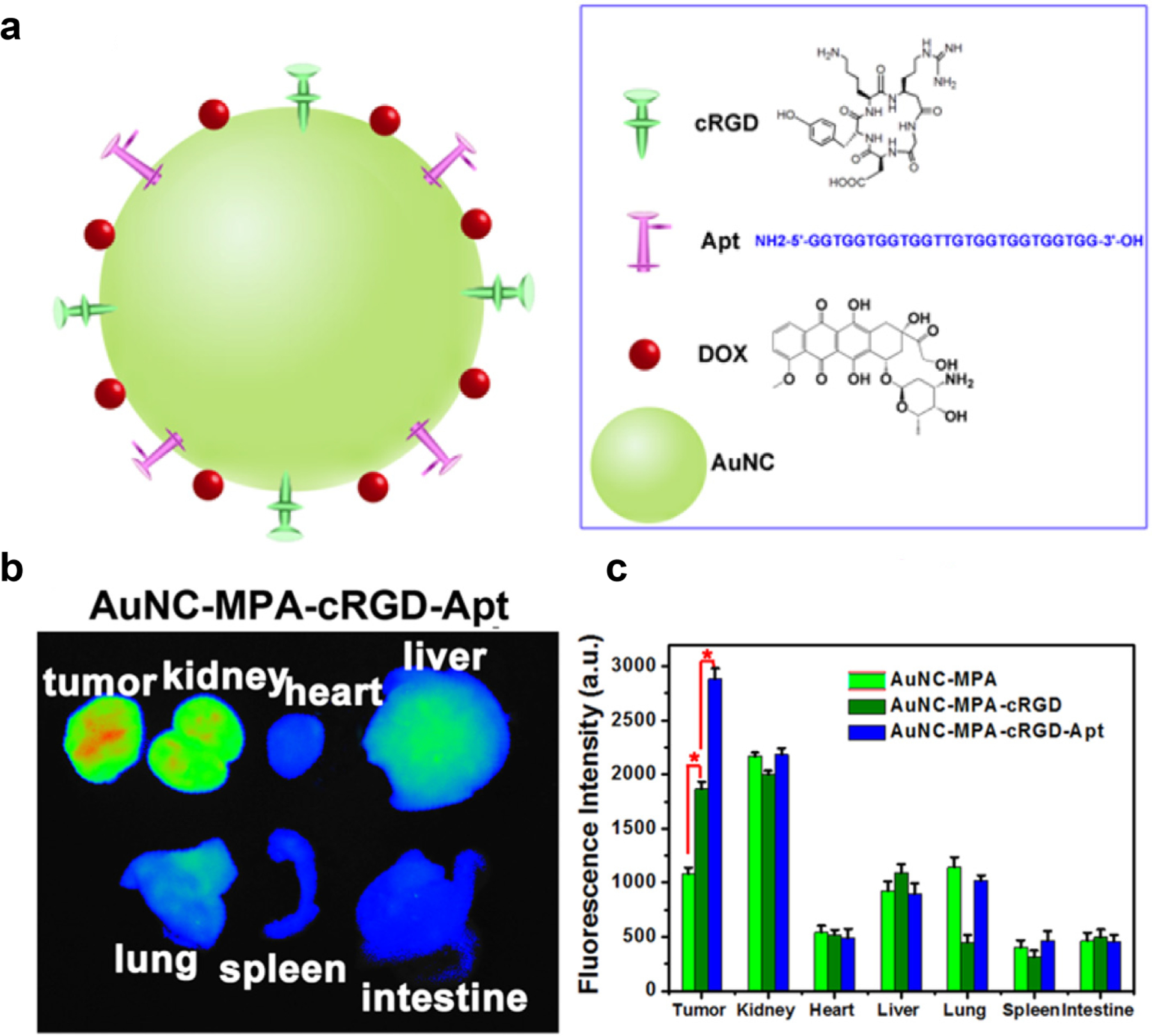
(a) Schematic structure of dox-loaded antibody/aptamer (cRGD/Apt)-conjugated AuNC. (b) ex vivo fluorescence images of isolated organs from tumor-bearing mice at 8 h post-injection. (c) Fluorescence intensity of isolated organs at 8 h post-injection with different sample formulations (Adapted from ref. 182).