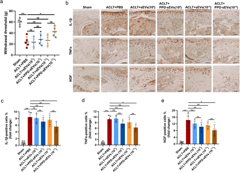
FIGURE 9.

In vivo evaluation of PPD‐sEVs for OA related pain in ACLT‐induced mice. (A) Mechanical sensitivity was measured after ACLT surgery (n = 5). WT, withdrawal threshold; (B) Representative images of immunohistochemistry analysis for pain‐related inflammatory cytokines including IL‐1β, TNFα, and NGF in each group, scale bar: 100 μm; (C‐E) Quantitative analysis of immunohistochemical staining of pain‐related inflammatory markers (n = 5), the fold change of each marker in each group was normalized against the Sham group (vs. Sham group). *P < 0.05, **P < 0.01
