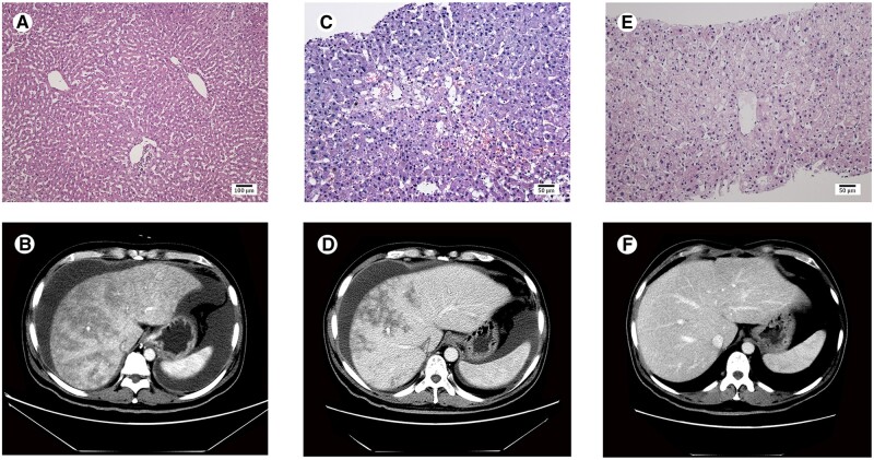
Figure 1.
Pathological manifestations and computed tomography imaging of the liver. Histological examination of the donated liver at the time of transplantation is normal (A). Liver biopsy shows significant dilation and congestion of the hepatic sinusoids in zone 3 of the hepatic acinus, regional hepatocellular necrosis, infiltration of the space of Disse by a few red blood cells, and deposition of collagenous fiber in the centrilobular veins on day 58 after transplantation (C) and recovered sinusoidal and centrilobular vein lesions on day 174 after transplantation (E). Abdominal computed tomography scan shows enlarged liver with patchy enhancement, massive ascites, and obscured hepatic veins on day 52 after transplantation (B), improved patchy enhancement, reduced ascites, and clear hepatic veins on day 90 after transplantation (D), and normal liver and hepatic veins without ascites on day 136 after transplantation (F). A, HE staining, ×100; C, HE staining, ×200; E, HE staining, ×200.

