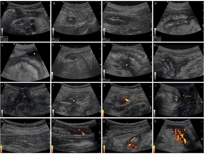
Figure 1.
Representative ultrasonographic findings. (A) Thickened terminal ileal wall (thickness: 4.9 mm) compared to cecal wall. (B) Thickened (5.7 mm) intestinal wall in transverse section and reactive hyperechoic mesentery (*). (C) Ulcer located in the affected wall showing transmural involvement. (D) Thickened (11 mm) terminal ileal wall with stratified involvement and mesenteric fibrofatty proliferation (*). (E) Reactive mesentery (*) around diseased terminal ileal segment with accompanying fluid (circle). (F) Prestenotic dilatation (double-headed arrow) proximal to the diseased segment (arrowheads). (G) Abscess (*) and related fistulae (arrows) originating from the diseased ileal segment with stratified involvement. (H) Fistulous tract between the terminal ileum and sigmoid colon (arrows). (I) Enterocutaneous fistulous tracts (arrows) with hyperechoic air inside. (J) Enteroenteric fistulae involving adjacent intestinal loops with center shrinkage (star sign). (K) Appendiceal involvement showing thickened terminal ileum and appendix with accompanying vascularity. (L) Transperineal ultrasound showing abscess (*) related to the fistulae (arrows) extending from the anal canal to the skin. (M) Limberg grade 1: Barely visible vascularity in the intestinal wall. (N) Limberg grade 2: Obvious vascularity as prominent vascular spots. (O) Limberg grade 3: Longer stretches of vascularity in the involved segment. (P) Limberg grade 4: Vascularity reaching mesentery.

