Figure 1.

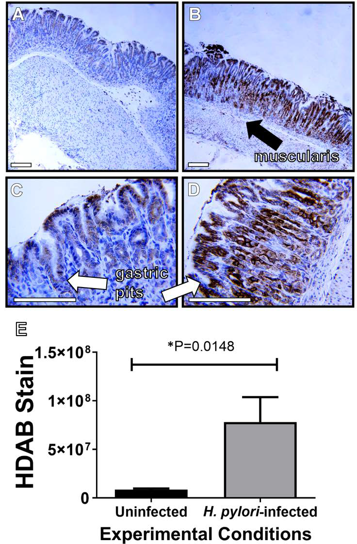
Lactoferrin is elevated in H. pylori-infected gastric tissues compared to uninfected gastric tissues. Immunohistochemical (IHC) and microscopical evaluation of gerbil gastric tissue specimens reveals elevated lactoferrin levels (indicated by the dark brown stain) associated with H. pylori chronic infection (B and D) compared to uninfected negative controls (A and C). Micrographs were collected at 100X (A and B) and 400X magnification (magnification bars indicate 100 μm) and representative micrographs are shown (N=3–4). Healthy, uninfected gastric tissue produces lower levels of lactoferrin, largely associated with gastric epithelia proximal to the lumen of the stomach. Infected tissues produce lactoferrin at higher levels and deeper into tissues towards gastric pits (white arrows) and muscularis (black arrows). H-DAB stain quantification (panel E) reveals that lactoferrin levels are significantly higher in H. pylori-infected gastric tissues compared to uninfected control tissues (*P=0.0148, Student’s t test with Welch’s correction).
