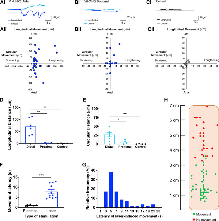
Figure 3.
Optogenetic stimulation of distal colon epithelium initiates change in local motility. Ai: representative traces illustrate the epithelium-evoked movement in the distal colon of Vil-ChR2 mice. Aii: movement patterns were characterized by tissue movement in the longitudinal and circular directions (n = 6 mice). Bi: example traces show smaller movement patterns in the proximal colon of Vil-ChR2 mice. Bii: movement patterns in the proximal colon were quantified in the longitudinal and circular directions (n = 4 mice). Ci: control littermate mice showed very minimal changes in local motility, demonstrated by the representative traces. Cii: quantification of longitudinal and circular movement also shows minimal movement in control colons (n = 4 mice). D: epithelial stimulation in the distal colon produced local movements in the longitudinal direction that were significantly greater than those produced by proximal colon epithelial stimulation (P = 0.002; one-way ANOVA) and in colons from control littermate mice (P = 0.002; one-way ANOVA). E: evoked movements in the circular direction were also significantly larger in the distal colon compared with proximal colon (P = 0.03; one-way ANOVA) and control colon (P = 0.006; one-way ANOVA). F: there was a latency between the start of blue light stimulation and colon movement which was significantly longer than the latency with electrical stimulation (P = 0.0005; Mann–Whitney test). G: latencies ranged from 3 to 22 s, with the majority of responses occurring within 6 s from the onset of laser. H: schematic of flattened colon preparation indicating where, in Vil-ChR2 mice, blue light stimulation of the epithelium initiated tissue movement above spontaneous baseline movement and where blue light stimulation failed to produce tissue movement. *P < 0.05, **P < 0.01, ***P 0.0005. ChR2, channel rhodopsin.

