
FIGURE 1.
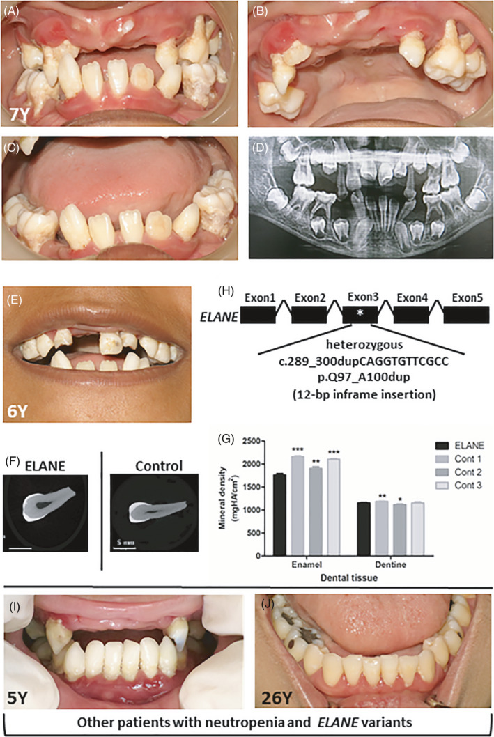
Oral phenotype and genetic mutation in a SCN patient. (A‐C) Oral photographs of the patient at 7 years old demonstrated severe periodontitis, dental infection, ulcers, dental caries, tooth mobility and premature tooth loss. (D) Panoramic radiograph of the patient demonstrated alveolar bone destruction, malocclusion, tooth extrusion and premature loss of deciduous teeth. (E) The photograph of the patient at 6 years old illustrated a deciduous upper left central incisor with enamel hypomineralization. (F) Micro‐computed tomography (Micro‐CT) images of the deciduous upper left central incisor of the patient and control. (G) Micro‐CT analysis revealed that the mineral density of the patient's tooth enamel was significantly less than that of controls. (H) The patient was identified with the heterozygous 12‐bp inframe insertion, c.289_300dupCAGGTGTTCGCC; p.Q97_A100dup, in ELANE. (I, J) The other two unrelated patients who had neutropenia and ELANE variants showed tooth infection, hypomineralized enamel and gingival inflammation