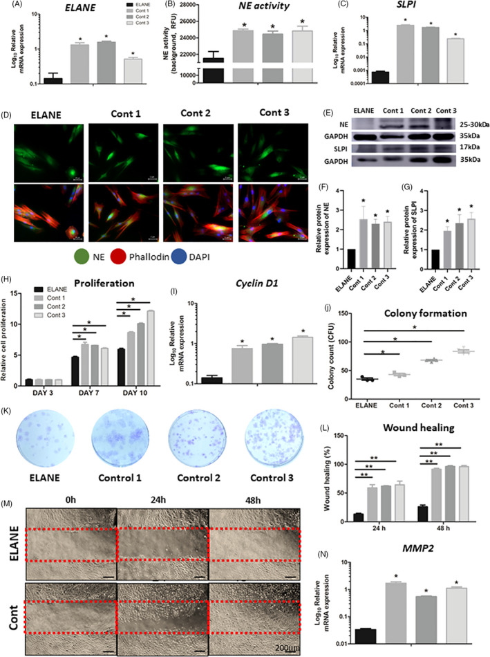
FIGURE 2.

Gene and protein expression, enzyme activity, proliferation, colony formation and wound healing. (A‐C) The mRNA expression of ELANE and SLPI, and neutrophil elastase (NE) activity of ELANE cells were significantly less than those of control. (D) Immunofluorescence detected NE staining in ELANE cells compared with controls. NE/ neutrophil elastase (green); Phalloidin (red); DAPI (blue). (E‐G) The NE and SLPI protein expression was significantly reduced in ELANE cells compared with that in controls. The GAPDH bands denote the controls for the NE or SLPI bands located above. (H) MTT assay showed that ELANE cells are significantly less proliferative than controls at Day 7 and Day 10. (I) The Cyclin D1 mRNA expression was significantly reduced in ELANE cells. (J, K) ELANE cells presented significantly less colony formation than controls. (L‐N) ELANE cells had delayed migration and wound healing and reduced MMP2 expression compared with controls. A significant difference between ELANE cells and controls: *p < 0.05; **p < 0.005; ***p < 0.0005. Cont, Control
