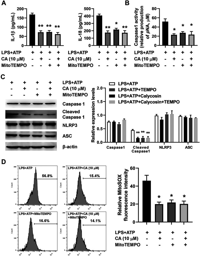
FIGURE 8.
Calycosin inhibits LPS-induced NLRP3 inflammasome activation through reducing mitochondrial ROS production in BMDMs. The LPS+ATP-induced BMDMs were treated with calycosin (10 μM) in the absence or presence of MitoTEMPO (10 μM) for 24 h. (A) The levels of IL-1β and IL-18 in the supernatant were determined by ELISA assay. (B) The caspase 1 activity in BMDMs was determined by a commercial kit. (C) The protein levels of NRLP3 inflammasome were determined by western blot. The quantitative densitometry of the protein expressions was determined by Image J software. Densitometric values were normalized to β-actin. (D) The LPS+ATP-induced BMDMs were treated with calycosin (10 μM) in the absence or presence of MitoTEMPO (10 μM) for 12 h. Cells were harvested and further incubated with MitoSOXTM Red (5 μM) for 10 min at 37°C in the darkness. Cell samples were acquired with a FACScan flow cytometer (Becton Dickinson, United States) with an excitation wavelength of 620 nm and an emission wavelength of 580 nm. Fluorescent signal intensity was analyzed by FlowJo software. Data are presented as means ± SEM. *p < 0.05; **p < 0.01 vs. the LPS+ATP group.

