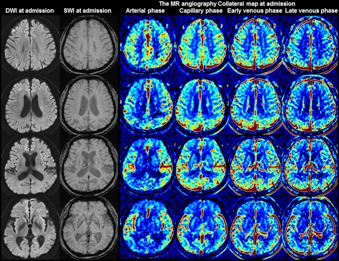
Figure 1.
Images of a 68-year-old man with greater than 70% stenosis of the right proximal internal carotid artery. The premorbid modified Rankin scale (mRS) score of this patient was 0, and the National Institutes of Health Stroke Scale score at admission was 1. The diffusion-weighted imaging (DWI), susceptibility-weighted imaging (SWI), and multiphase MR angiography collateral map at 44 min after symptom onset are shown. DWI shows acute infarct signals along the border zones of the right cerebral hemisphere, and SWI shows no prominent cortical and medullary veins in the right cerebral hemisphere, representing good collateral status. The MR angiography collateral map shows no collateral-perfusion delay in the right cerebral hemisphere in the capillary phase (MR acute ischemic stroke collateral score of 5: excellent collateral perfusion defined as no or small collateral-perfusion delay in the ischemic MCA territory in the capillary phase regardless of the collateral status in the arterial phase). The patient underwent conservative treatment and recovered, as shown by the 90-day mRS score of 0.

