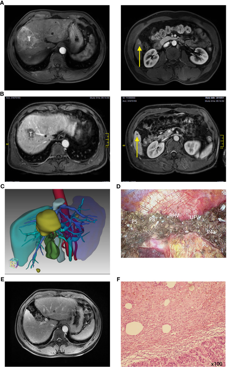
Figure 3.

A typical case had the combination therapy and underwent subsequent surgery (case no. 6). (A) Radiology of case no. 6 before treatment: T for tumor 1 and yellow arrow pointing tumor 2. (B) Radiology of case no. 6 after treatment: T for tumor 1 and yellow arrow pointing tumor 2. (C) Three-dimensional reconstruction and surgical plan of case no. 6 after the combination therapy. (D) Surgical photo of the margin (P for pedicle, RHV for right hepatic vein, and UFV for umbilical fissure vein). (E) Reexamination of the liver 6 months after the surgery: No tumor or relapse. (F) Pathology of tumor: moderately differentiated hepatocellular carcinoma (HCC) is focally distributed with extensive necrosis and several inflammatory cells (×100); viable tumor cell rate, 2.5%.
