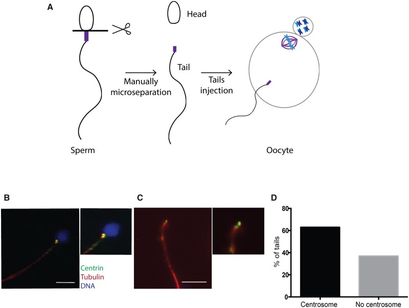
Figure 5.
The sperm basal body localizes to the microsurgically separated tails. (A) Schematic representation of our functional assay. (B) IF for centrin, tubulin and DNA on intact sperm. Scale: 7.5 μm. (C) Representative IF images of a manually separated sperm tail stained for centrin, tubulin and DNA. Scale: 7.5 μm. (D) Graph showing the percentage of isolated tails with centrosomes. N = 2 different sperm samples.

