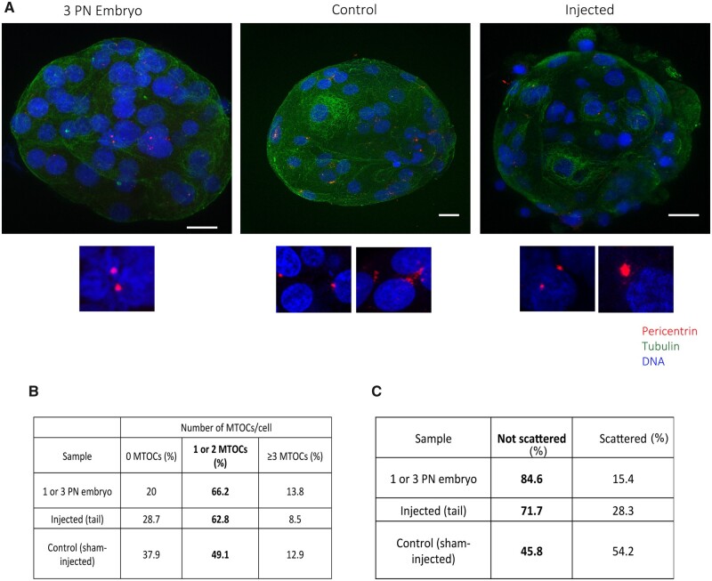
Figure 7.
Microtubule organizing centers (MTOCs) can be formed de novo in control pseudo-blastocysts and after the activation of the embryonic genome. (A) Representative IF images of blastocyst and pseudo-blastocyst of abnormal fertilized oocytes, control and injected oocytes stained for pericentrin, tubulin and DNA. The lower panels are magnifications of the MTOCs for each condition. Scale: 20 μm. (B) Number of MTOCs per cell in abnormal fertilized oocytes, control and injected oocytes. (C) Table showing the percentage of scattered MTOCs per sample. N = 5 3PN embryos, N = 10 control and N = 15 injected oocytes.

