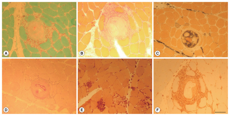
Fig. 1.
Biopsy of the quadriceps muscle. Gomori trichrome stain (A) and Hematoxylin & Eosin stain (B) reveal cysts surrounded by inflammatory cells. The material encapsulated in the cysts stain with Alkaline Phosphatase (C) and larvae stain strongly with Periodic Acid Schiff (PAS) staining (D). Necrotic muscle fibers are sporadically observed, as well as muscle fibers replaced by large histiocytic cells stained with Esterase staining (E). Inflammatory cells surrounding cysts and invading muscle fibers are macrophages CD68 immunopositive (F). Scale bar in (F) 100 μm (valid for (A–F)).

