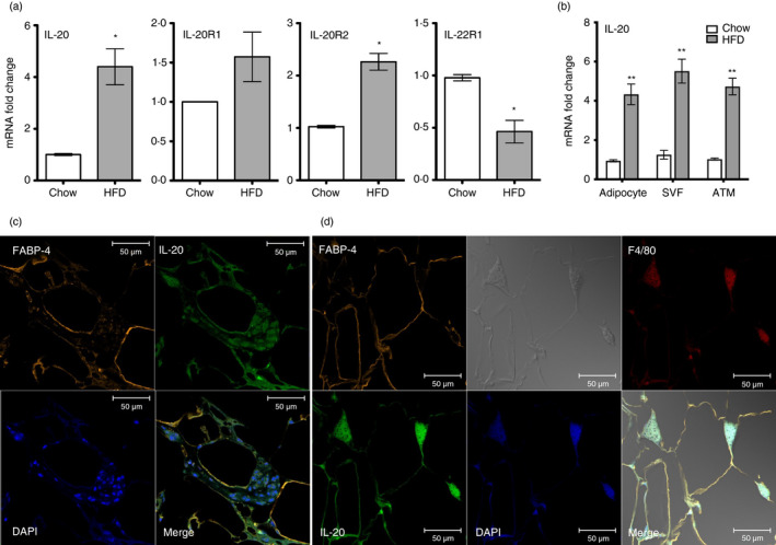
FIGURE 2.

IL‐20 was upregulated in visceral white adipose tissue and macrophages in obese mice. (a) RT‐qPCR analysis of IL‐20, IL‐20R1, IL‐20R2 and IL‐22R1 mRNA expression changes in the visceral white adipose tissue (WAT) of HFD mice (n = 6) for 16 weeks and LFD mice (n = 6) for 16 weeks. (b) Relative mRNA expression of IL‐20 in the adipocytes and stromal vascular fractions (SVFs) isolated from the visceral WAT of LFD mice (n = 5) and HFD mice (n = 5) for 16 weeks. (c) Immunofluorescence staining for the adipocyte markers FABP‐4 (orange), IL‐20 (green) and DAPI (blue) in the visceral WAT of HFD mice. Co‐localization of IL‐20 with FABP‐4 is shown in yellow in the merged image. Scale bars =50 μm. (d) Immunofluorescence staining for the adipocyte markers FABP‐4 (orange), IL‐20 (green), the macrophage marker F4/80 (red) and DAPI (blue) in the visceral WAT of HFD mice. Co‐localization of IL‐20 with FABP‐4 and F4/80 is shown in yellow in the merged image. Scale bars =50 μm. Data in (a) and (b) are expressed as mean ±SEM *p < 0·05, **p < 0·01. Data are representative of three independent experiments
